Mineralized Tissue Histology
The histologic tissue processing, photography and microscopic imaging area houses equipment for the processing of mineralized and unmineralized tissues for microscopic examination. There is a dedicated space for dissection, photography, and microscopy.
Mineralized Tissue Histology

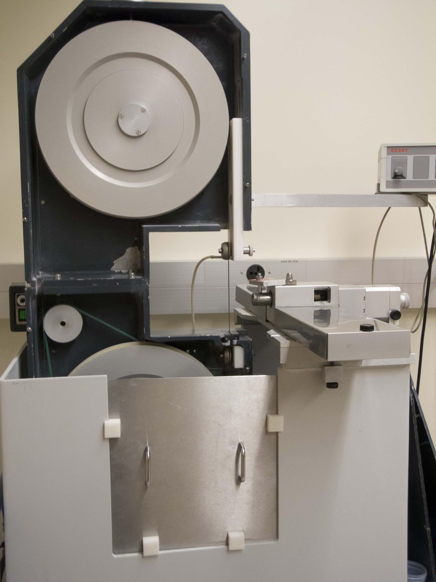
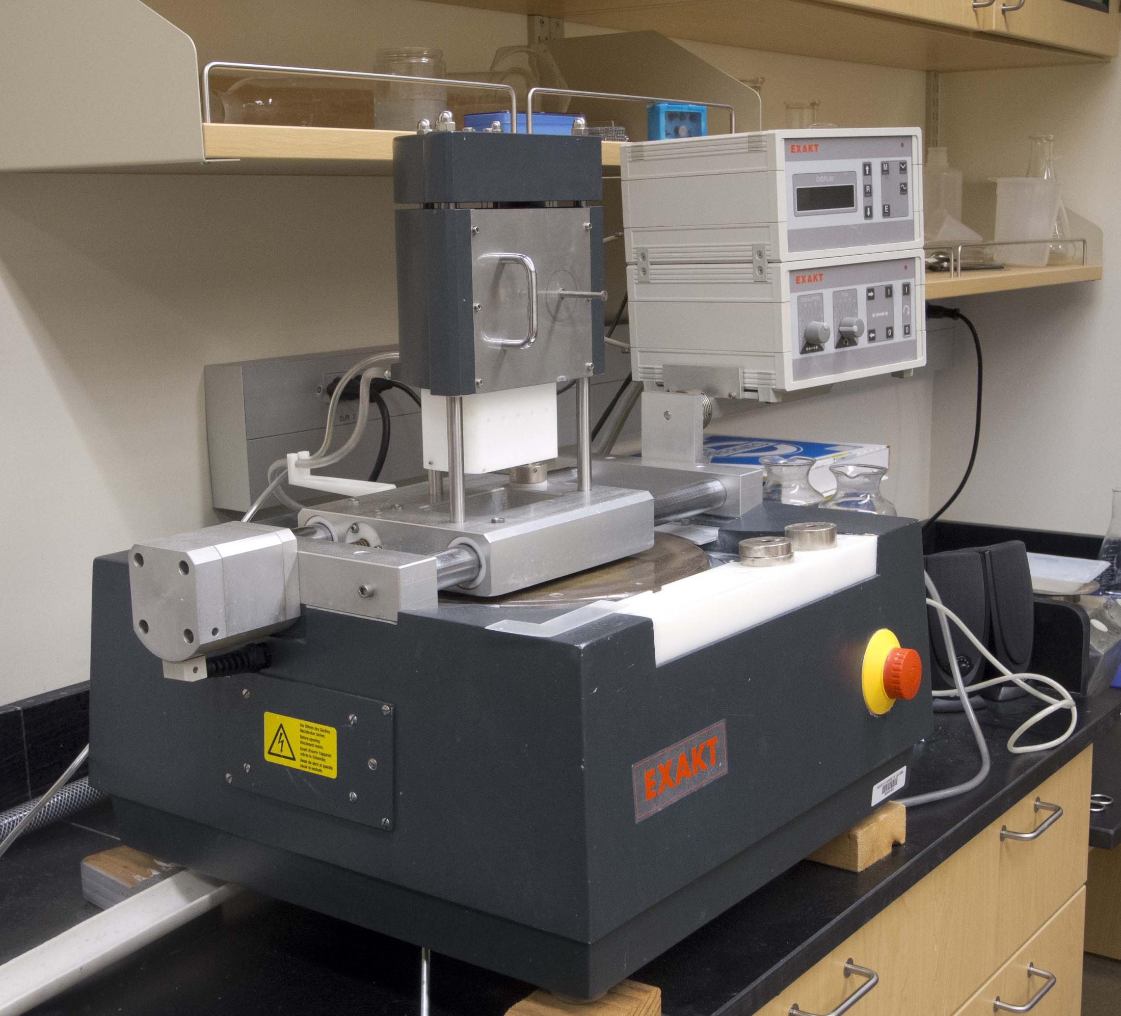
The tissue processing area houses equipment for the plastic embedment and processing of mineralized (and unmineralized) tissues for microscopic examination. This equipment includes a Leitz 1600 circular bone saw, a Buehler Isomet low speed bone saw, a Buehler Ecomet 2 grinder/polisher, a Maruto laper, a Polycut E microtome/ultramiller, and an Exakt 310 CP System. The Exakt Cutting/Grinding System is an integrated system for sectioning undecalcified tissues and metallic implants that consists of a precision band saw, embedding tools, a microgrinding device, and accessories for specimen handling.

Microscopy
The microscopy area houses an Olympus Vanox II Research Photomicroscope with transmitted light, epifluorescent, phase contrast, polarized light, and Nomarsky capabilities. It has a PAXcam and PAX-It software for image capture, measurement, and processing. An Olympus stereophotomicroscope is also available. Custom software allows for general morphometry or for quantitative microradiography.
Photography
The photography area houses a photostand and Nikon D3 SLR digital camera, Nikon D1X SLR digital camera, Nikon Coolpix P7100 digital camera, Nikon 990 digital camera, and SONY Handycam digital video camera for documentation of tests and findings.
