Facilities
The J.D. Wheat Veterinary Orthopedic Research Laboratory is an organized research center of the School of Veterinary Medicine at the University of California at Davis. The multi-user laboratory in building VetMed3A consists of a multi-room cell & molecular biology area (3000 sq ft), mechanical testing facility and fabrication workshop (2800 sq ft), small animal motion-capture/gait lab (1575 sq ft), medical models fabrication/3D printing area (600 sq ft), mineralized tissue processing area (605 sq ft), histology processing area (200 sq ft), microradiography area (260 sq ft), microCT specimen scanner (240 sq ft), photography areas (90 sq. ft.), microscopy area (90 sq ft), computer area (650 sq ft), and graduate student, staff and faculty office space (1500 sq ft) in Vet Med 3A. Much of the equipment and measurement techniques are provided as a service on a recharge basis (see Laboratory Services). The laboratory is adjacent to the Veterinary Medical Teaching Hospital and California Animal Health and Food Safety Laboratory which facilitates collaboration.
Cellular and Molecular Biology
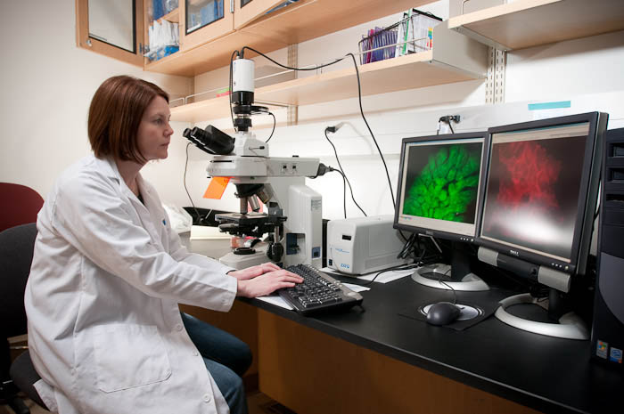
The cell biology laboratory is fully equipped for cell culture and routine biochemical and molecular biology procedures. The microscopy area houses a state of the art microspectrofluorometry system (inverted NIKON Eclipse TE 2000-U microscope with epifluorescence and a Sutter filter switcher), equipped for quantifying cytosolic ion concentration, fluorescent dye transfer and image analysis using Metamorph.
Computing
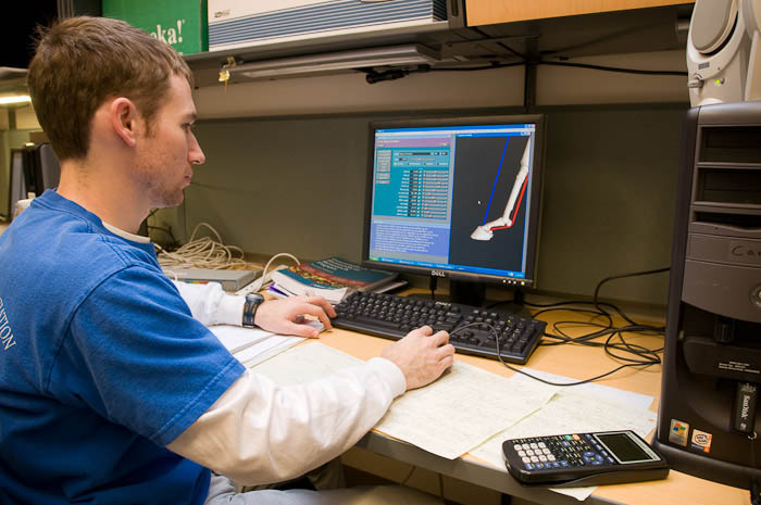
The computer room houses 5 PC computers running Windows 10 or 11 and the student/staff workrooms house 5 PCs and 3 iMacs. Various computers are loaded with Word, Excel, SigmaPlot, Powerpoint, Access, SAS, Egret, Photoshop, ImageJ, Motus, FlexTestGT, MultiPurpose Testware, Labview, SIMM, SD Fast, Dynamics Pipeline, Matlab, Visual C++, Fusion360, and ProE, Dragonfly and Osirix, Abaqus, Mimics, 3Matic, Visual 3D, and internet software.
Gait and Locomotion
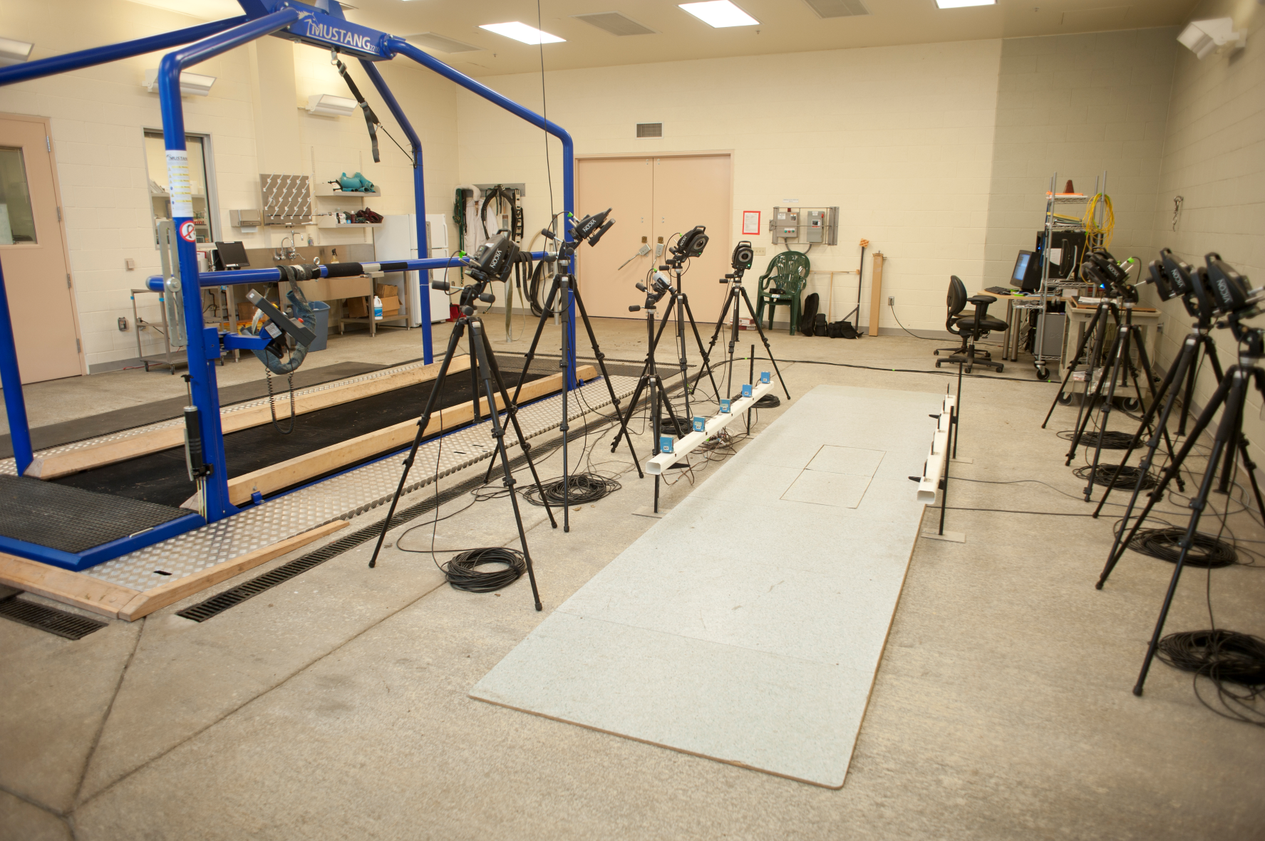
Kinetic, motion capture, and pressure systems are housed in 3 rooms in the Equine Athletic Performance Building. Kinematic and kinetic data acquisition systems are configured for companion and equine animals, and for research studies. A 3-D kinematic data acquisition and analysis system including six Qualisys Miqus optical cameras and 2 color view cameras is synchronized with dual portable Kistler force plates configure for canine use. Weight-bearing pressure and force can be acquired using the 3-tile Tekscan Strideway walkway. Standing weight-bearing for canines is measured with a StanceAnalyzer system.
Mechanical Testing
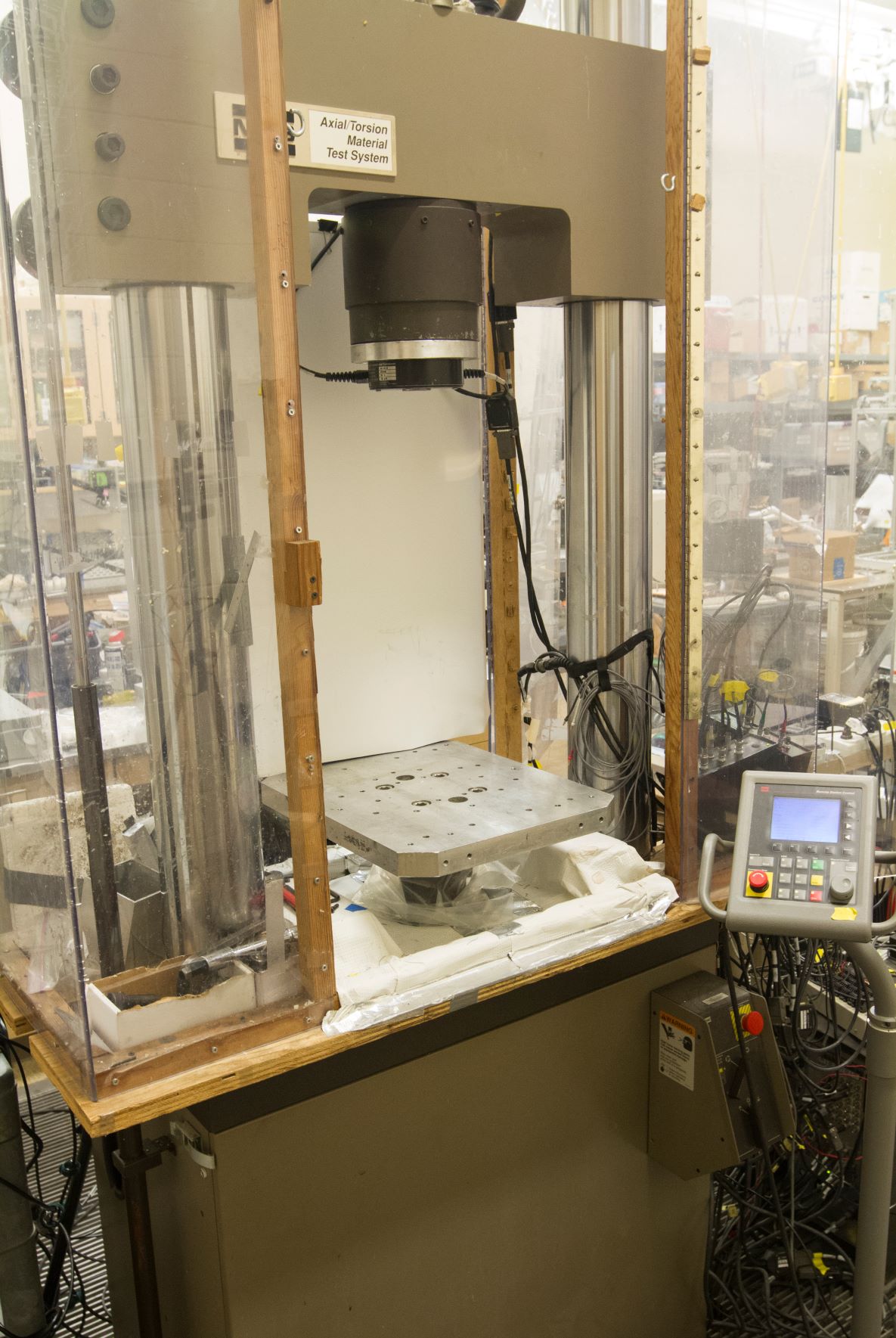
The mechanical testing facility houses a Materials Testing System Model 809 axial-torsional servohydraulic mechanical testing system capable of generating 55,000 lb axial load and 25,000 in-lb torsional load. For medium to small specimens, there is an Instron 5960 single-axis electromechanical system with a 1kN and 50N load cell. For very small specimens and for specialized cartilage indentation mapping or hardness indentation mapping for mechanical properties, there is a Biomomentum Mach-1 with multiaxis 90 N load cell and a 15 N load cell.
Medical Modeling and Manufacturing
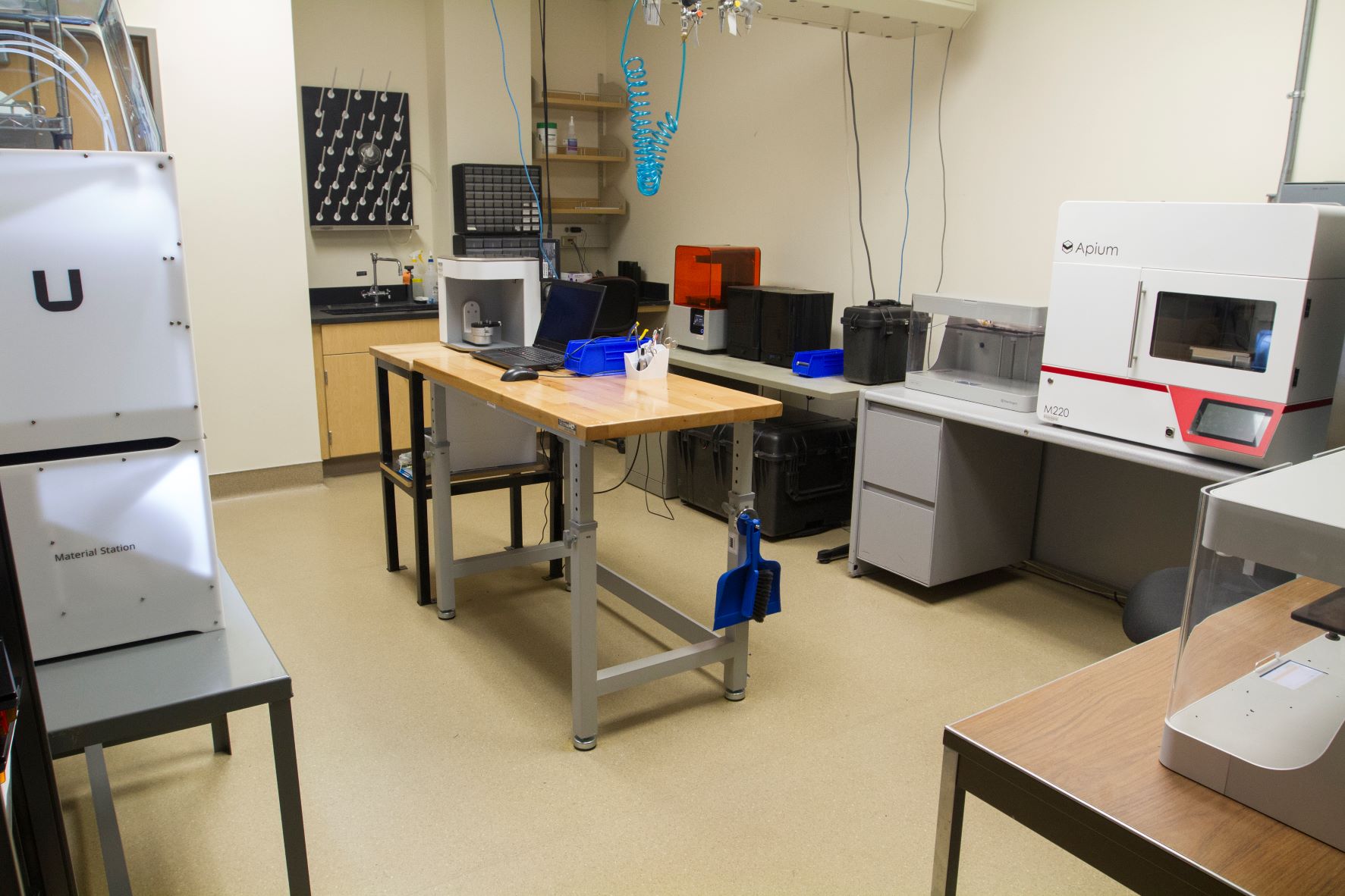
The MMM area contains 2 Markforged printers (Onyx), an Ultimaker dual-head printer (multi-materials), an Apium medical PEEK printer and a Formlabs SLA printer, and a Artec Micro high-resolution 3D scanner. The MMM area also includes a dedicated space for artificial bone production and a machine shop that includes a Tormach CNC mill, bandsaw, drill presses and grinder.
Mineralized Tissue Histology
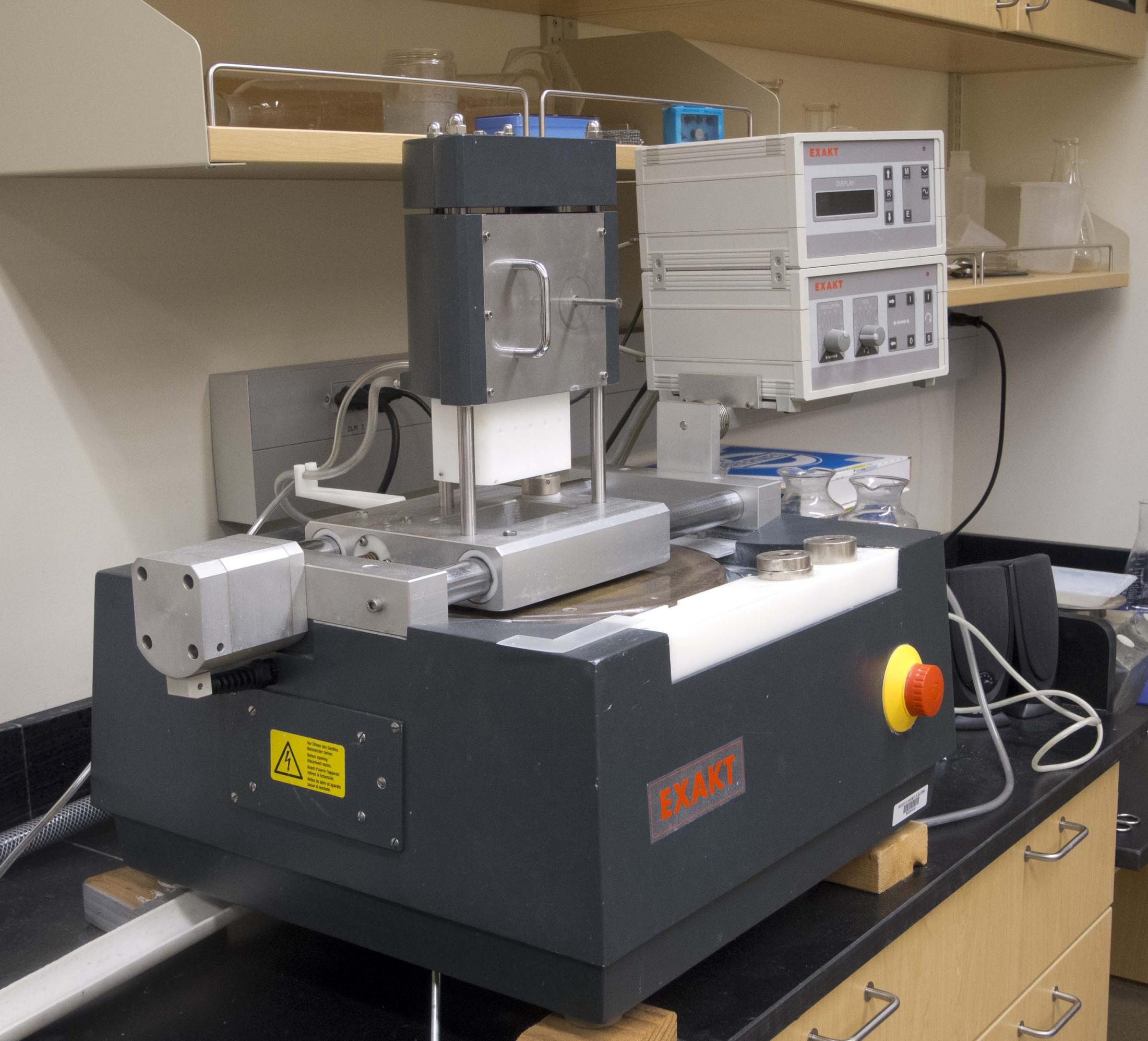
Mineralized Tissue Histology
Microscopy
Photography
Musculoskeletal Imaging
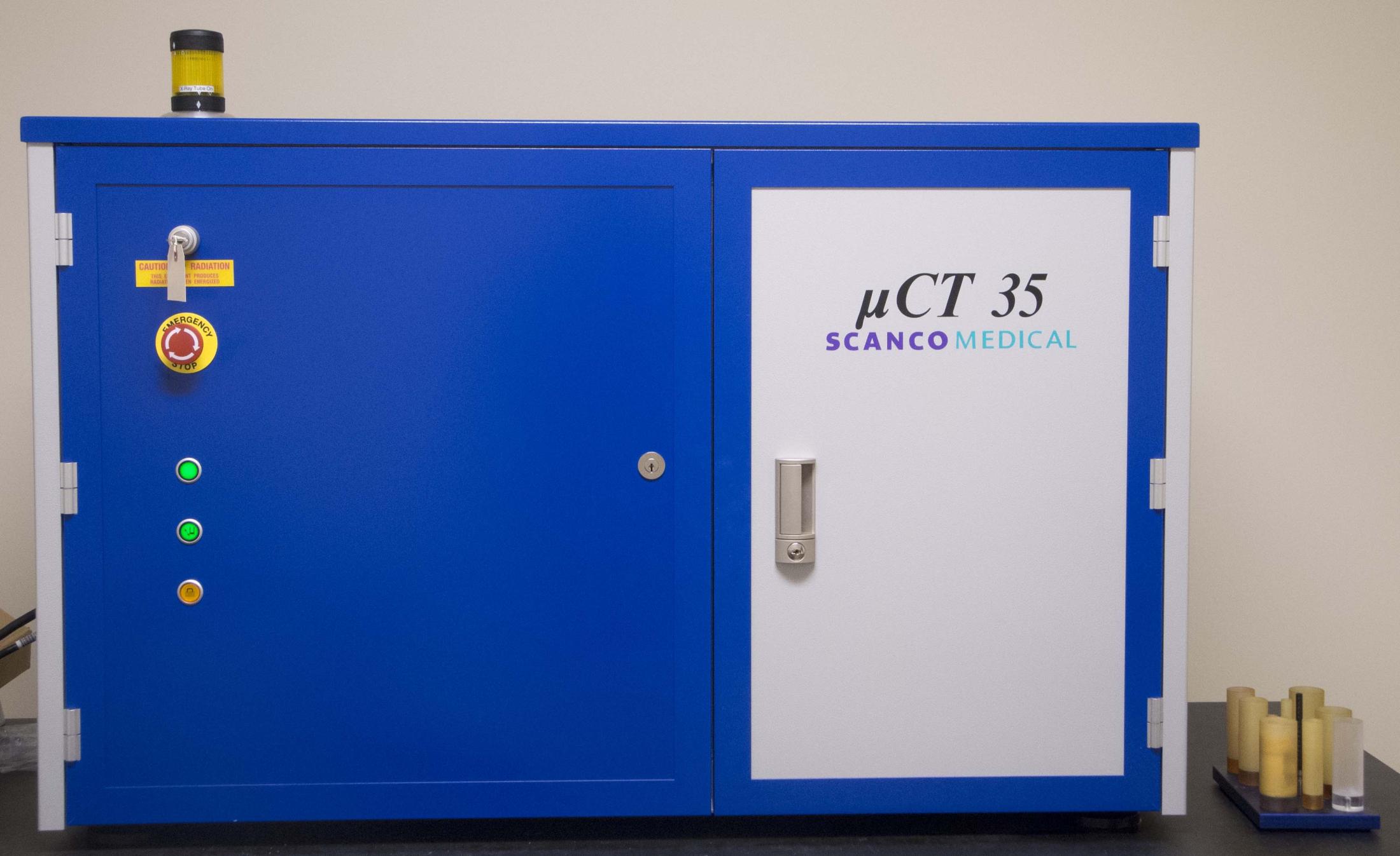
The microCT area houses a ScanCo Medical μCT35 specimen scanner. Its capabilities are ex-vivo specimens from size 35mm diameter by 74mm long (37 micron resolution) to 6mm diameter by 45mm long (5 micron resolution). It is runs a 45-70kVp, 72-145 microAmp conebeam. It includes calibration of CT image to Hydroxyapetite for bone analysis and software to analyze density, bone volume, and morphometry measures. It has the capability of scanning soft-tissues using contrast-enhancement (blood vessels, cartilage, etc).
